Outlining what MRI misses
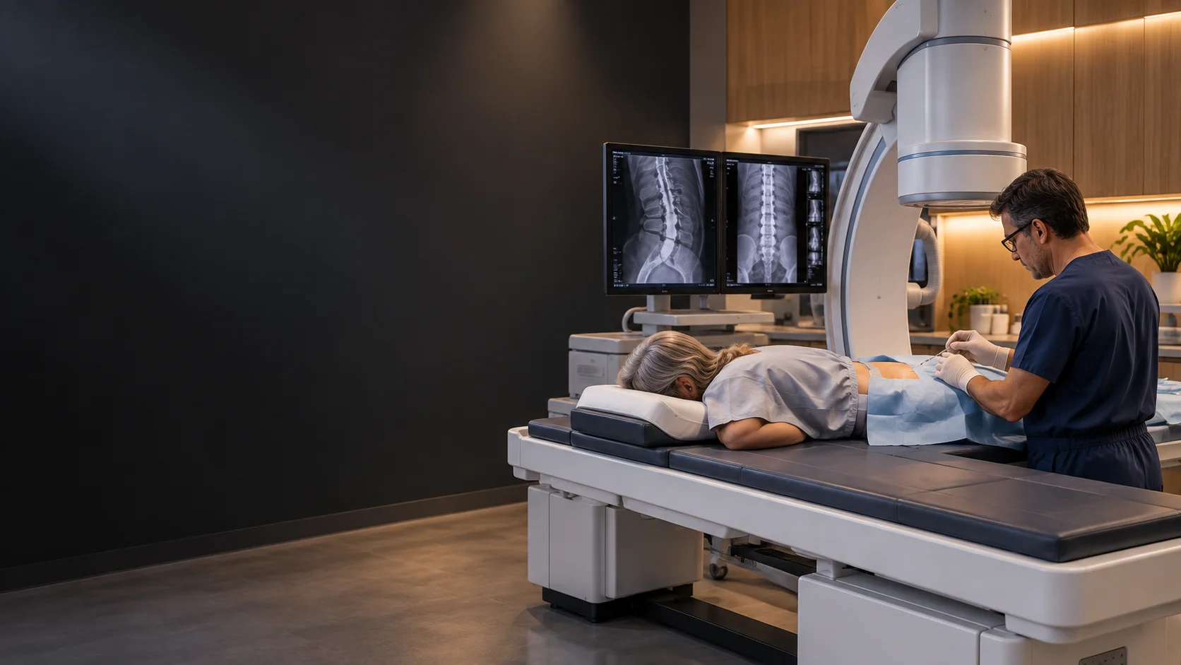
Contrast dye placed in the spinal sac coats the cord and nerve roots, and CT slices then show exactly where the outline narrows or deforms.
Diagnostics · Invasive
An imaging study of the spinal canal using contrast dye to outline the cord and nerves.
What it is
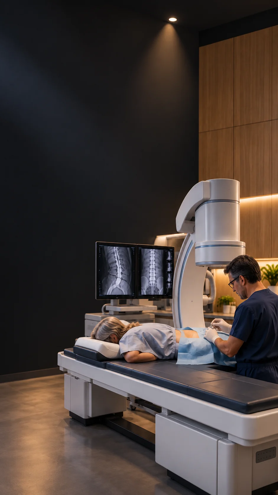
Contrast dye placed in the spinal sac coats the cord and nerve roots, and CT slices then show exactly where the outline narrows or deforms.
Typically reserved for cases where MRI is inconclusive, contraindicated by implants, or degraded by previous metalwork — or to confirm a level before surgery.
Because images can be taken in different positions, myelography can reveal narrowing that appears only under load or movement.
It involves a spinal puncture, so it is used only when the answer genuinely changes the plan — never as routine.
Common questions
MRI images tissue directly and non-invasively. Myelography images the fluid space around the cord with contrast — useful precisely where MRI falls short, such as around metal implants.
The skin and deeper tissues are numbed first. Most people feel pressure rather than pain, plus a brief headache risk the team explains beforehand.
Blood thinners, infections, seizure history, kidney disease, allergies and pregnancy all matter. Bring a full medication list so the team can plan safely.
You rest flat for a period and drink fluids while the contrast clears. The team gives exact aftercare instructions for your case.
Free · No obligation · Reviewed by the ProSpine team