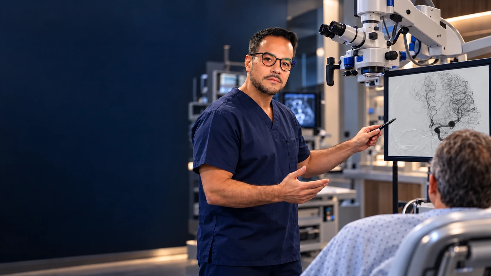
The operating microscope
Magnification plus coaxial light reveals tissue planes, tiny vessels and nerve fibres invisible to the naked eye.
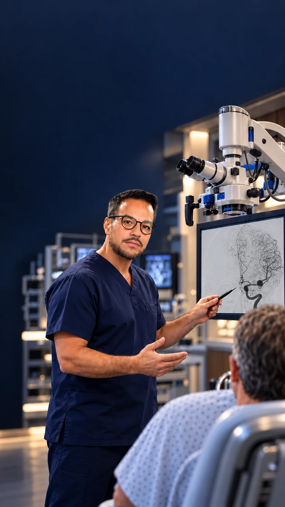
Treatments · Brain Surgery
Microsurgical and vascular procedures of the brain and spine, performed within the full neurosurgical scope of the practice.
What it is
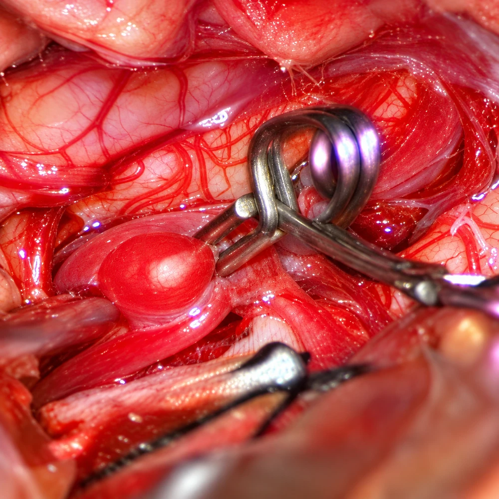
Magnification plus coaxial light reveals tissue planes, tiny vessels and nerve fibres invisible to the naked eye.
Aneurysms, malformations and cavernomas demand control of blood flow measured in millimetres — the microscope's home territory.
Distinguishing lesion from healthy brain or cord depends on seeing the interface, which only magnification provides.
Smaller, exact corridors mean less retraction of healthy tissue on the way in and out.
What to expect
Approach, positioning and each operative step are defined from imaging before the day of surgery.
Where the cord or critical pathways are at stake, electrophysiological monitoring watches function continuously.
Neurological observation leads early recovery, with rehabilitation starting from whatever baseline surgery preserves. Report fever, wound changes or new neurology promptly.
Common questions
The routine use of the operating microscope for dissection, vessel handling and closure — technique and magnification, not a separate specialty.
Brain and cord tumours, vascular malformations, and any lesion wrapped around critical nerves or vessels — your imaging decides.
Bleeding, stroke, infection and new neurological deficit are the honest risks of operating near eloquent tissue; monitoring and planning exist to minimise them.
A multidisciplinary review of your imaging and symptoms — surgery only when it is genuinely the right path for your condition and goals.
Free · No obligation · Reviewed by the ProSpine team